

Dr Norman Marcus
Modern medicine relies heavily on imaging, laboratory testing, and advanced technologies. For many patients with chronic pain, the most revealing diagnostic tool is still a careful, hands-on evaluation of the body’s muscles.
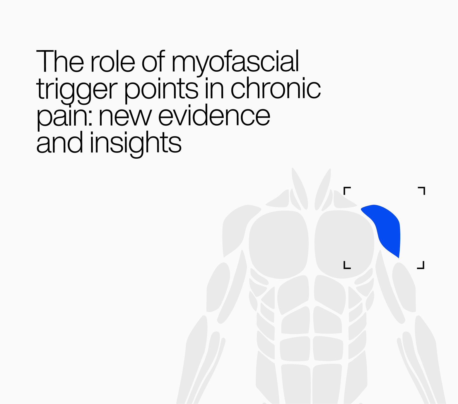
Muscles represent the largest functional system in the human body and play a central role in posture, stability, and movement. Increasingly, research shows they are also frequent contributors to chronic pain conditions, particularly in the back, neck, shoulders, and hips.
Despite this clinical relevance, structured training in muscular examination remains limited in many medical programs. When muscular assessment is overlooked, clinicians may rely primarily on imaging or structural explanations, sometimes leaving the true source of pain unaddressed.
A thorough muscle examination is therefore an essential component of accurate pain diagnosis. Below are practical steps clinicians can use to evaluate muscular contributions to chronic pain.
A meaningful diagnosis starts with understanding how the pain behaves. Muscle pain is often activity-dependent, positional, or triggered by specific movements.
Key questions include:
Patterns of onset and aggravation frequently reveal muscular origins. Pain that increases with sustained posture or improves with gentle movement often reflects muscle-related dysfunction rather than fixed structural pathology.
Before touching a patient, a physician can gather powerful clues simply by watching them stand, sit, bend, or walk.
Look for:
Movement observation often reveals compensations or overload patterns that develop when certain muscles are weak, fatigued, or overactive.
This is the most critical and most overlooked step.
Palpation enables clinicians to identify:
A skilled palpation of muscles such as the quadratus lumborum, paraspinals, gluteal muscles, piriformis, trapezius, and hip flexors can reveal pain generators that imaging will never detect.
Muscles fail in multiple ways. Some are weak; others are tight; many are simply unable to perform repeated tasks without fatiguing quickly.
Doctors should evaluate:
These findings often differentiate muscle-driven pain from nerve or joint pathology.
Referred pain is one of the defining features of muscle dysfunction. A trigger point in the gluteus medius can mimic sciatica; tension in the lumbar paraspinals can create flank or lower abdominal pain; tight cervical muscles can produce headaches.
Doctors should compare a patient’s symptoms against well-documented myofascial referral maps. Recognizing these predictable referral patterns helps distinguish muscular pain from other conditions when imaging findings are inconclusive.
A simple but powerful test:
Pain that appears or worsens during a specific muscle contraction strongly suggests a muscular origin.
Some emerging diagnostic approaches use controlled electrical stimulation to identify muscle fibers that respond abnormally to activation. While not yet routine in all clinical settings, this method can help clarify uncertain cases by highlighting muscles that generate disproportionate pain responses.
Muscle examination is not a relic of traditional medicine but a clinically valuable skill that complements modern diagnostics. When muscular assessment becomes part of routine evaluation, clinicians gain a clearer understanding of how pain develops and persists.
Integrating careful observation, palpation, and functional testing allows physicians to identify muscular sources of pain earlier and tailor treatments more effectively.
In many cases, a thoughtful physical examination can reveal what technology alone cannot.
Muscles matter and careful clinical examination remains one of the most powerful tools for understanding pain.